Total Pageviews

Monday, September 28, 2020

Genetic testing in autism


What is the risk of having a second child with autism? It is interesting to know that it is higher (7%) if the first child with autism was a girl than if it was a boy ( 4%). If a couple has 2 children with autism, the risk of having yet autistic child shoots up to 33-50%.


The genetic architecture of autism is complicated. Largely it is due to the presence of 2 or more low or moderate risk variants which are difficult to identify.https://dx.doi.org/10.1590%2FS1679-45082017RB4020

About 10% will have an abnormal chromosomal microarray. Clinical clues are dysmorphism, malformations & a family history of psychiatric disorders.

There are just a handful of monogenic disorders which are associated with autism.

The most important would be the Fragile X syndrome. It may be wise to recommend it in males with autism even in the absence of the classical phenotype and in girls who have a family history of fragile X or premature ovarian failure.

Girls with intellectual delay and autism need to be tested for the MECP2 gene.

Children with a large head > 2.5 SD need evaluation for the PTEN mutation.

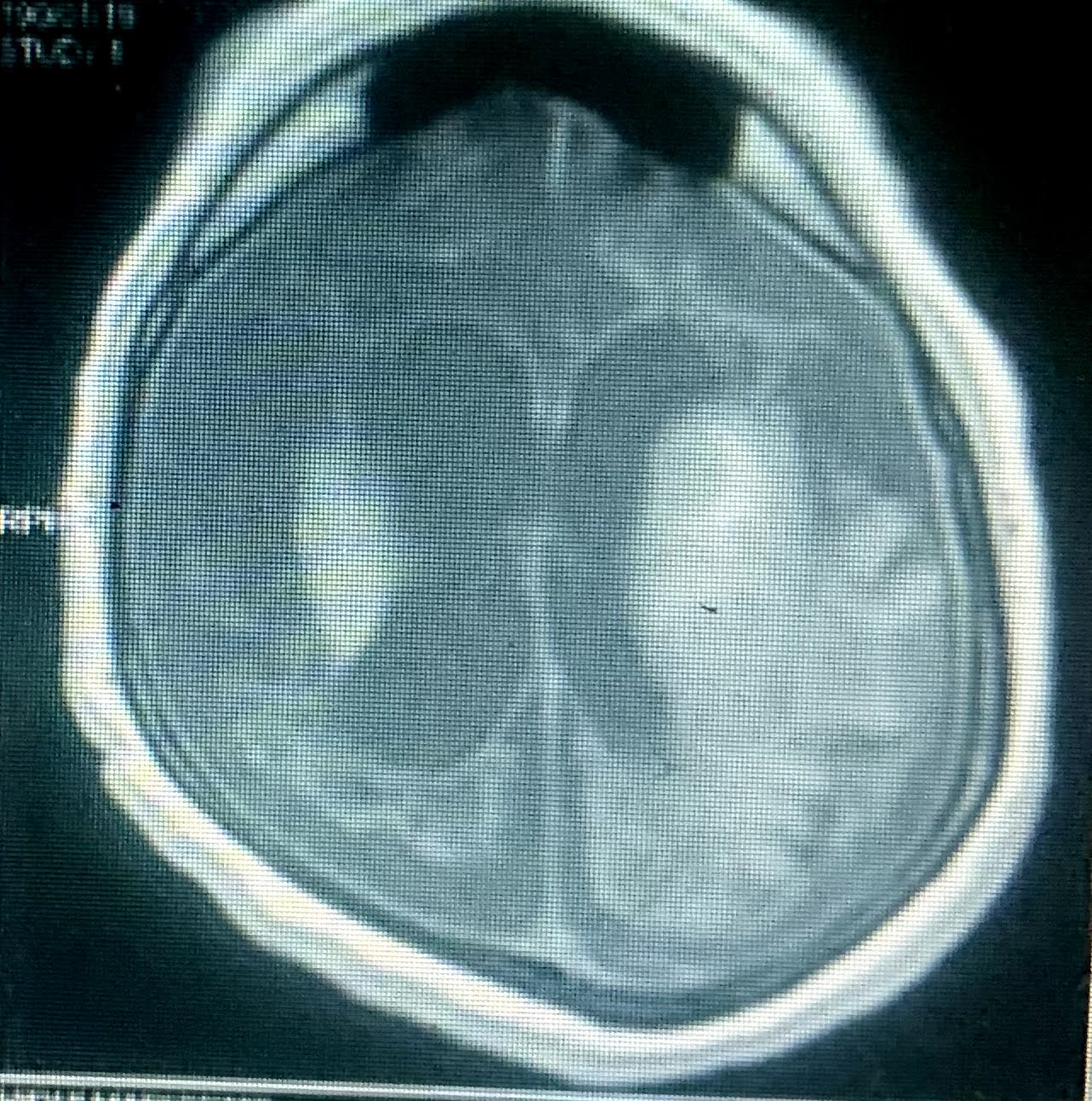
If there are seizures, episodic deteriorations you may need to rule out IEM's. Some treatable IEM's like creatine deficiency disorders may be picked up on an MR spectroscopy.


The bottomline is that no single genetic test will make a diagnosis of autism. It is a clinical diagnosis but about 25% may have an underlying genetic disorder.


Sunday, September 20, 2020

GBS associated with COVID 19 - what should we know?


This 5 year old girl came to our hospital with fever and non specific myalgia 8 days back followed 3 days later by progressive ascending quadriparesis.

Her nerve conduction velocities were suggestive of an acute motor axonal neuropathy. However her nasopharyngeal swab for COVID 19 was positive.

What must we know about GBS associated with COVID 19?

Caress et al have reviewed 37 cases of GBS associated with COVID 19 published in literature so far.https://doi.org/10.1002/mus.27024


The time to onset of neurological symptoms after COVID 19 ranged from 3-28 days ( mean 11 days). It appears to be slightly more rapid than usual.

The vast majority (84%) were symptomatic with primary COVID symptoms during the GBS.

Interestingly facial diplegia was seen in almost a quarter of patients.

The requirement for ventilation was 37.8% slightly higher than the routine 20-30% in the garden variety of GBS. It may to some extent be explainable because of the associated respiratory involvement in SARS COV2 infection.

CSF PCR for SARS CoV 2 was negative in all the 18 in whom it was tested so a direct viral damage seems unlikely.

The COVID 19 virus attaches via the spike protein to the ACE 2 receptors and also to gangliosides containing silica acid residues.

So the hypothesis is that anti COVID antibodies in our bodies may inadvertently attack the gangliosides resulting in GBS. However antiganglioside antibodies have been seen in only 12% of the patients in whom they were tested.

 Treatment has been mostly IVIG or PLEX. One must be careful with IVIG since their is an underlying hypercoagulable state in SARS CoV-2 infections. Most people would avoid PLEX because of the possibility of hemodynamic compromise.

The risk of GBS with the COVID19 vaccine also needs to be monitored.


Sunday, September 13, 2020

The biochemistry behind Infantile Tremor Syndrome


A 9 month old baby came in with progressive hyperpigmentation for 15 days, tremors and altered sensorium for 5 days.

His haemoglobin was 7.7gm/dl with an MCV of 96fl. His MRI brain showed a predominant fronto-temporal atrophy and some mild delay in myelination. His Serum Vitamin B12 was low and serum homocysteine was elevated.


No marks for making the diagnosis of Infantile Tremor Syndrome (ITS). He had been inadequately weaned and was predominantly on breast feeds. His mother was a strict vegetarian. All this tied in very well with the classical picture of ITS. The clouds of doubt about the underlying ethology are slowly dissipating in the face of strong evidence that the culprit is vitamin B12 deficiency.

Studies from PGI Chandigarh in 92 children have shown that though Vitamin B12 levels are documented to be low in about 42%, serum homocysteine is a better marker and is elevated in 96.5 %. An MCV > 95fl is again seen in a mere 42 %.https://doi.org/10.1007/s12098-019-03117-w

If you have inadvertently done an  acylcarnitine profile you may pick up elevated C3/C2 and C3/C16 levels. Urine organic acids will show elevated methylmalonic acid levels.

How does cobalamin deficiency result in such widespread neurological deficits in the brain?

Cobalamine in the brain is in 2 forms - adenosylcobalamine and methylcobalamin. Both have a role to play.

Adenosyl cobalamin converts methylmalonyl CoA  to succinylcholine CoA. In its absence excess of propionyl CoA is formed and subsequently odd chain fatty acids. These C15, C17 fatty acids when incorporated into the myelin are defective. Hence the abnormal myelination and developmental delay.


Methycobalamin deficiency on the other hand impairs conversion of homocysteine to methionine which in turn is the precursor of an important methyl donor for various fatty acids in myelin.


Tuesday, September 8, 2020

Does the herpes virus persist in the brain after encephalitis?


An 8 year old girl with cerebral palsy with spastic quadriparesis was admitted with the history of fever, seizures and altered sensorium. This was her MRI brain (seen above)with cystic encephalomalacia and fresh diffusion restriction in bilateral basal ganglia. Her CSF had 7 lymphocytes, elevated proteins and PCR for Herpes was positive with 4000 copies/ml.

In her neonatal period also she had been diagnosed with viral encephalitis and had been treated with acyclovir for presumed herpes infection on the basis of diffusion restriction of bilateral temporal lobes, which is seen the the MRI below. Despite treatment she had been left with significant neurological deficit and had become essentially bedridden.


There have been reports of herpes reactivation several years after an initial encephalitis. In fact it is believed that the virus often remains in a latent state in some tissues in the brain such as the ependymal lining of the ventricles even after treatment with standard doses of acyclovir.

In fact 70% of cases of HSV encephalitis is considered due to reactivation of the virus, though primary infections are more common in children.

During the latent period only the "Latency Associated Transcript" (LAT)of the virus is coded in the neurons. This also interestingly enhances neuronal survival by inhibiting apoptosis. During periods of immune depression such as infections or fever or even trauma, the virus is reactivated and enters the lytic cycle and starts actively damaging neutrons. https://doi.org/10.3389/fncel.2019.00046


There are also cases which have documented a progressive downhill neurological disease due to herpes encephalitis. It is also implicated in certain neurodegenerative disorders such as Alzheimers disease and multiple sclerosis. It is proposed that multiple mild HSV infections involving the brain may result in the same pathology noted in Alzheimers disease.

Acyclovir inhibits active viral replication. It has been suggested that viruses which are not actively replicating may continue to survive in the brain. Various suggestions including longer duration of treatment with acyclovir and repeated courses have ben suggested to overcome the problem of relapsing or chronic HSV brain disease.


Sunday, August 30, 2020

What links congenital CMV, Aicardi-Goutiere's Syndrome and Non Megalencephalic Cystic Leukoencephalopathy?


A 3 year old boy came in with the history of neuroregression for the past 6 months. His head size was normal.

His MRI brain was striking. He had cysts in bilateral temporal lobes and multifocal sub cortical white matter changes in frontal and parietal-occipital lobes. 

It decided to relook at all the important leukodystrophies with cysts.

1. Megalencephalic Leukoencephalopathy with subcortical cysts- This has diffuse, extensive white matter changes. The white matter looks positively swollen. The child is usually developing normally and may develop brief deterioration after minor head trauma. In short the MRI brain looks much worse than the child. It's extremely common among the Agarwal community in India. Most importantly they all have a large head which my patient lacked.

2. Congenital CMV disease- Congenital CMV disease also presents with white matter changes and temporal cysts. They often have cortical malformations like polymicrogyria, ventriculomegaly, calcifications and cerebellar atrophy. They are symptomatic since early infancy and have microcephaly.

3. Aicardi Gautiere syndrome- They mimic congenital CMV. They present after a few weeks of life with irritability and sterile pyrexia and chillbalians. The basal ganglia calcifications are striking besides the temporal cysts and white matter changes. 

4. Cystic Leukoencephalopathy without megalencephaly- These children are symptomatic from birth with global developmental delay and very slow to static clinical progression of disease. The head size is normal and MRI brain shows temporal cysts and scattered multifocal subcortical white matte changes, which our patient had.

Congenital CMV, AGS and cystic Leukoencephalopathy without megalencephaly are called interferronopathies.


Normally viral nucleic acids trigger release of interferon. Interferon production can also be upregulated by the presence of excessive nucleic acid residues derived from what are called retro elements. These are remnants of ancient viruses in our DNA which are usually metabolised by various enzymes coded by genes like TREX1, RNAase H2 etc. Mutations in these genes result in excessive nucleic acid residues which activate interferon and cause this group of auto inflammatory disorders like Aicardi Gautieres and cystic Leukoencephalopathy without megalencephaly.  https://doi.org/10.1111/dmcn.14268

This is the key to the uncanny similarity between congenital CMV and AGS.


Sunday, August 23, 2020

Undeciphering the mTOR pathway and the Indian scientist who started it all.


This 8 year old boy came in with the history of developmental delay and epilepsy. The diagnosis was written on his face. 

The adenoma sebaceum which you see is a misnomer for what are actually angiofibromas. They are a classical skin manifestation of tuberous sclerosis (TS).

Tuberous sclerosis is the classic mTORopathy. Dysregulation in the mTOR pathway is the reason behind the various manifestations including cardiac rhabdomyomas, intracranial hamartomas, subependymal giant astrocytomas and renal angiolipomas.

Overactivation of the mTOR pathway results in giant dysplastic neutrons, abnormal axonogenesis and dendrites, disordered cortical lamination reduced myelination and increased excitatory synaptic currents.

This mechanism has also been implicated in disorders with cortical malformations due to other genes such as STRADa, DEPDC5 and PI3K which act upstream of the mTOR pathway.


Since then there are been great excitement about possible targeted therapy with mTOR inhibitors in TS.

The EXIST-3 was a phase 3 RCT which used Everolimus (an mTOR inhibitor) and showed significant reduction of seizures in patients with TS ( 14.9% in placebo, 29.3% in low dose and 39.6% in high dose group).https://doi.org/10.1016/S0140-6736(16)31419-2

However a recent RCT everolimus failed to improve cognition/ autistic features in children 4-7 years old with TS.


I sat down to understand the mTOR pathway and was pleasantly surprised to find that it all began due to the exemplary work of an Indian scientist Surendra Nath Sehgal. http://www.indianjcancer.com/article.asp?issn=0019-509X;year=2017;volume=54;issue=4;spage=697;epage=698;aulast=Samanta

Surendra Nath Sehgal studied pharmacology from BHU, India; went on to do a PhD from Bristol and finally settled down in a research lab in Canada. He isolated a drug from the soil sample of the exotic Easter Islands in the South Pacific Ocean. 

Easter Island, Rapa Nui, Moai, Statue

He called it Rapamycin because the island was called Rapa Nui.

Though initially evaluated as an anti-fungal it was subsequently found to have immunosuppressive actions.

His persistent efforts to further characterize its properties led to the discovery of its significant anti cancer effects. Incidentally, I suppose that's why the commonest side effect of the mTOR inhibitors like everolimus is stomatitis.

Intense research into its mechanisms revealed that it acts on a target (to be later called mTOR) which plays a key role in cell proliferation and cell growth. This pathway is switched on by many oncogenic signalling pathways.

Ironically, Surendra Sehgal developed metastatic colon cancer in 1998 and he was treated with Rapamune which helped him survive till 2003.
 

Sunday, August 16, 2020

Allan-Herndon-Dudley Syndrome: a medical chimera

 A 10 month old boy came to me with the history of axial hypotonia, scissoring of lower limbs and dyskinetic movements for the past 6 months. 

His birth weight was 3.75 kg but despite a good appetite had poor weight gain and excessive sweating. His parents also noticed that the anterior fontanelle had closed by 6 months. 

His TSH was normal but he had an elevated fT3 and normal fT4. His free T3/T4 ratio was 4.13

This combination of developmental delay with dyskinesias in a boy with a free T3/T4 > 0.75 is classical of   Allan-Herndon-Dudley syndrome (AHDS).


The problem in AHDS is in the MCT8 gene. It codes for a protein which transports thyroid hormones across the blood brain barrier. In its absence T3 is unable to enter the brain and just a minuscule amount of T4 enters via another transporter coded by OATP1C1.

There is an excess of peripheral conversion of T4 to T3. What results is a peripheral thyrotoxicosis. But alas the brain is starving for thyroid hormones.

This lack of thyroid hormone inside the brain results in hypomyelination, developmental delay and dyskinesias. Systemically there is tachycardia, sweating and weight loss.

I sat down to see what is new in the therapy of this fascinating disease.

Merely giving T4 supplements aggravates the hyperthyroidism and is to be avoided.

Trials of a combination of T4 with propylthiouracil (PTU) stabilises the peripheral hyperthyroidism, allows a little T4 to enter the brain and improves weight gain. Unfortunately it does not improve the neurological functions significantly. Its drawback is the long term risk of agranulocytosis and liver failure with PTU.

A recent review of advances in therapeutics of AHDS describes the use of T3 analogs which don't require MCT8 to cross the BBB. Trials are on with three drugs- DITPA, TRIAC and sobetirome.

Work is also on with gene therapy and pharmacological chaperones ( Frontiers in Neurology April 2020) .https://doi.org/10.3389/fnins.2020.00380

An interesting study used intranasal T4 to bypass the block due to MCT8 deficiency but failed to raise thyroid hormone levels in the brain (PLOS ONE July 2020). https://doi.org/10.1371/journal.pone.0236113


Living on the edge- watershed infarcts

  A 3 year old boy with disseminated staphylococcal disease was referred to our hospital in  severe sepsis. He was in multi organ dysfunctio...